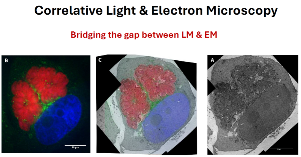
Hsia. et. al. 2015 Studying Membrane Trafficking in Toxoplasma gondii Using Correlative Light and Electron Microscopy (CLEM)
Correlative light and electron microscopy (CLEM) is a powerful technique to study molecular and structural interrelationships. It combines two well-established imaging techniques whereby researchers can, for the same specimen, take advantage of imaging a relatively large sample size by light microscopy and of the high resolution power of the electron microscope. CLEM makes it possible to target rare and/or dynamic events for structural analysis at high resolution.
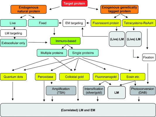
A significant advantage of CLEM is its versatility. For instance, CLEM can be combined with live cell imaging and 3D EM volume analysis to name two heavily used, state-of-the-art imaging approaches. The design of a CLEM experiment is often dependent on and can be tailored to the size, location and abundance of the target protein, the types of markers and antibody probes, and the aim of the project. The figure below outlines some of the possible workflows for a CLEM project. Please contact EML to schedule a consultation if you are interested in exploiting CLEM approaches in your research.

Fig 1. Flowchart of the methods for correlative LM and EM described in this chapter. These include
photo oxidation, enzymatic-based detection, and particle-based methods. See text for detaik.
Markers for Correlated Light and Electron Microscopy, Sosinsky et al. Methods in Cell Biology Vol 79