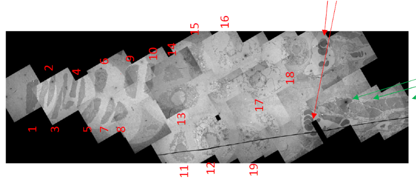
Human diseases, e.g., genetic, infectious, malignant or metabolic, often result in the manifestation of disorders at multiple levels. Improvements in diagnosis, treatment and prevention of diseases, as well as the study and characterization of disease-associated pathologies often requires the examination of large and complex tissue areas at both nanometer and micrometer scales. This is particularly true when examining a specific region of interest (ROI) at the sub-organelle level of resolution and a specimen in the millimeter(s) range. To accomplish this task, EML staff first generate a low magnification map using a semi-automated multi-frame tiling and stitching method, then zoom in and search for the specific ROI at higher magnification. This labor-intensive process is akin to searching for a needle in a haystack! The first image is a bone growth plate manually stitched together from 50 individual transmission electron microscope (TEM) images.

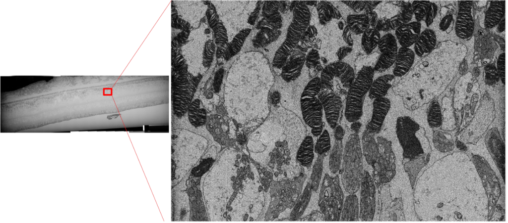
The recent development of high-resolution field emission scanning EM (SEM) with automated scanning navigation program has enabled multiscale imaging by first scanning a large piece of resin-embedded tissue using automation. The specific ROI can be then selected and re-imaged at higher resolution. The example below is a 1mm length retina tissue that was first scanned at low resolution. The specific ROI was then selected, and images were obtained at higher magnification to reveal ultrastructural details. This innovation has proven to be an invaluable time saving technology and will be an essential tool for all the biological EM core facilities.
