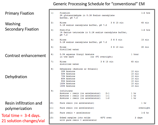
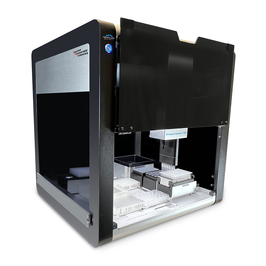
Conventional Electron Microscopy (EM) sample processing for tissue and cells was developed in the sixties. Sixty years later, similar sample processing workflows continue to be followed by many EM laboratories. These conventional workflows consist of more than 20 steps involving solution exchange and the handling hazardous reagents. Up until recently, most of these procedures were performed manually and often required two to three days to complete (Figure 1). Owing to these factors, EM sample processing is prone to variations. It is also difficult to apply bioEM techniques for high throughput screening. Recent studies have demonstrated that efficient mixing can shorten incubation time required for solution exchange and thus shorten the EM sample processing time. Furthermore, programmable robotic liquid handling systems are widely available and can readily be adapted to precisely reproduce EM sample processing steps. EML recently acquired a third generation automated EM sample processor, the PrepMaster 5100 (Figure 2). This instrument offers turnkey operation and can efficiently complete EM sample processing up to resin infiltration in 2 to 3 hours. Once programed, it only requires ~30 min of staff time to set up the solutions and the required lab ware.
EML is currently working on expanding the capability of the Prepmaster to process vibrotome or cryostat sectioned tissue slices, cells grown on coverslips, and also to perform immunolabeling experiments. Please contact EML if you are interested in adapting the Prepmaster to other automated experimental procedures.

