Transmission Electron Microscopy (TEM)
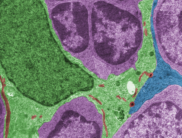
(TEM) is a microscopy technique in which a beam of electrons is transmitted through a specimen to form an image. The specimen is most often an ultrathin section less than 100 nm thick or a suspension on a grid. An image is formed from the interaction of the electrons with the sample as the beam is transmitted through the specimen. The image is then magnified and focused onto an imaging device, such as a fluorescent screen, a layer of photographic film, or a detector such as a scintillator attached to a charge-coupled device or a direct electron detector.


Scanning Electron Microscopy (SEM)
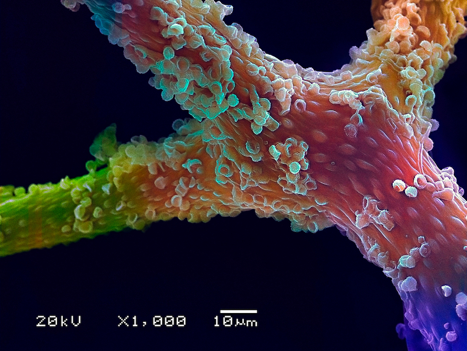
(SEM) is a type of electron microscope that produces images of a sample by scanning the surface with a focused beam of electrons. The electrons interact with atoms in the sample, producing various signals that contain information about the surface topography and composition of the sample. The electron beam is scanned in a raster scan pattern, and the position of the beam is combined with the intensity of the detected signal to produce an image. In the most common SEM mode, secondary electrons emitted by atoms excited by the electron beam are detected using a secondary electron detector (Everhart–Thornley detector). The number of secondary electrons that can be detected, and thus the signal intensity, depends, among other things, on specimen topography. Some SEMs can achieve resolutions better than 1 nanometer.
Immuno Electron Microscopy (IEM)
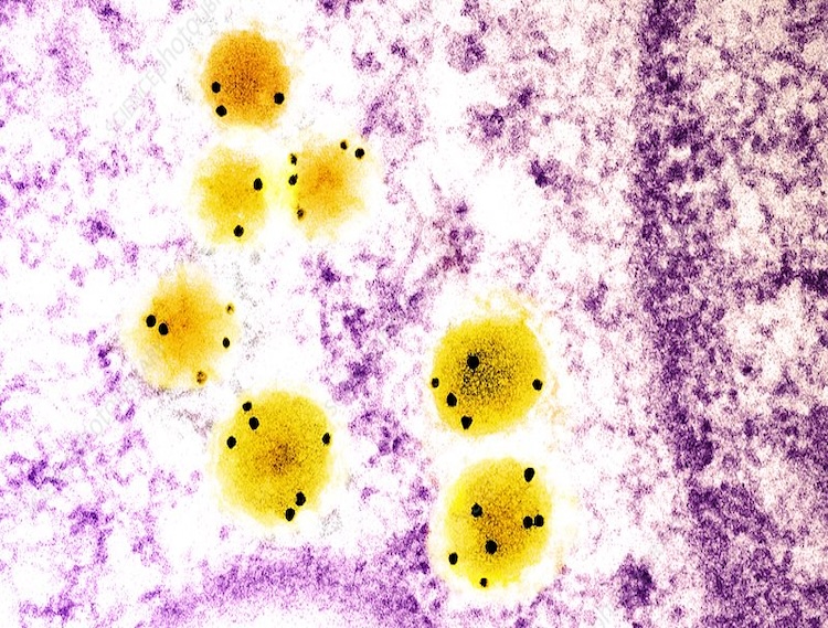
Immune electron microscopy (more often called immunoelectron microscopy) is the equivalent of immunofluorescence, but it uses electron microscopy rather than light microscopy. Immunoelectron microscopy identifies and localizes a molecule of interest, specifically a protein of interest, by attaching it to a particular antibody. This bond can form before or after embedding the cells into slides. A reaction occurs between the antigen and antibody, causing this label to become visible under the microscope. Scanning electron microscopy is a viable option if the antigen is on the surface of the cell, but transmission electron microscopy may be needed to see the label if the antigen is within the cell.


Negative Stain Transmission Electron Microscopy
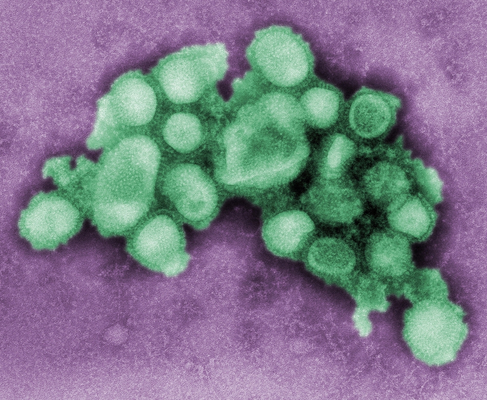
In the case of transmission electron microscopy, opaqueness to electrons is related to the atomic number, i.e., the number of protons. Some suitable negative stains include ammonium molybdate, uranyl acetate, uranyl formate, phosphotungstic acid, osmium tetroxide, osmium ferricyanide and auroglucothionate. These have been chosen because they scatter electrons strongly and adsorb to biological matter well. The structures which can be negatively stained are much smaller than those studied with the light microscope. Here, the method is used to view viruses, bacteria, bacterial flagella, biological membrane structures and proteins or protein aggregates, which all have a low electron-scattering power. Some stains, such as osmium tetroxide and osmium ferricyanide, are very chemically active. As strong oxidants, they cross-link lipids mainly by reacting with unsaturated carbon-carbon bonds, and thereby both fix biological membranes in place in tissue samples and simultaneously stain them.