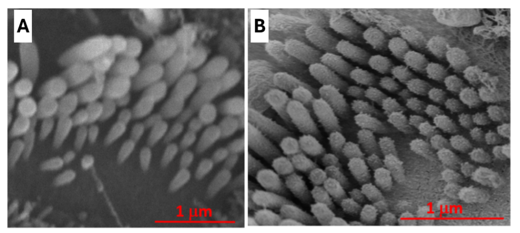
Traditional scanning electron microscopes (SEMs) have limited resolution when imaging biological specimens. Non-conductive biological samples need to be dehydrated and coated with a thin layer of conductive metal to prevent a charging effect and damage caused by the electron beam during imaging. The figure below illustrates the resolution of hair cells using traditional SEM (A) vs. FE-SEM (B). The tips of the hair cells can only be revealed when imaged using FE-SEM.

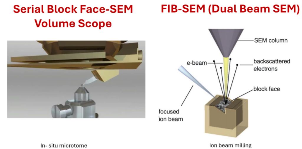
Modern FE-SEM is equipped with sensitive in-lens detectors and anti-charging mitigation strategies. Thus, it is also capable of conducting low voltage imaging of osmicated and resin embedded tissue specimens reaching resolution of ~ 2 to 5 nm, like traditional TEM. With advances in navigation and mapping software, FE-SEM enables pre-programmed imaging of large sample areas with automation. Furthermore, the capabilities of FE-SEM can be expanded by installing an in situ ultramicrotome (serial block face), additional focused ion beam (dual beam), or cryo chamber (cryo SEM). Below are schematic drawings that illustrate the configuration of a serial block face SEM and dual beam SEM. FE-SEM is a versatile and multifunctional instrument that soon will be a must-have instrument for all biological EM facilities.
