Instrumentation
At CCR volume Electron Microscopy Core, our primary techniques are focused ion beam scanning electron microscopy (FIB-SEM), array tomography (AT) and TEM electron tomography (ET), often used in conjunction with correlative light microscopy, to image um- to mm-sized samples in 3D and at nanoscale resolutions, at room temperature.
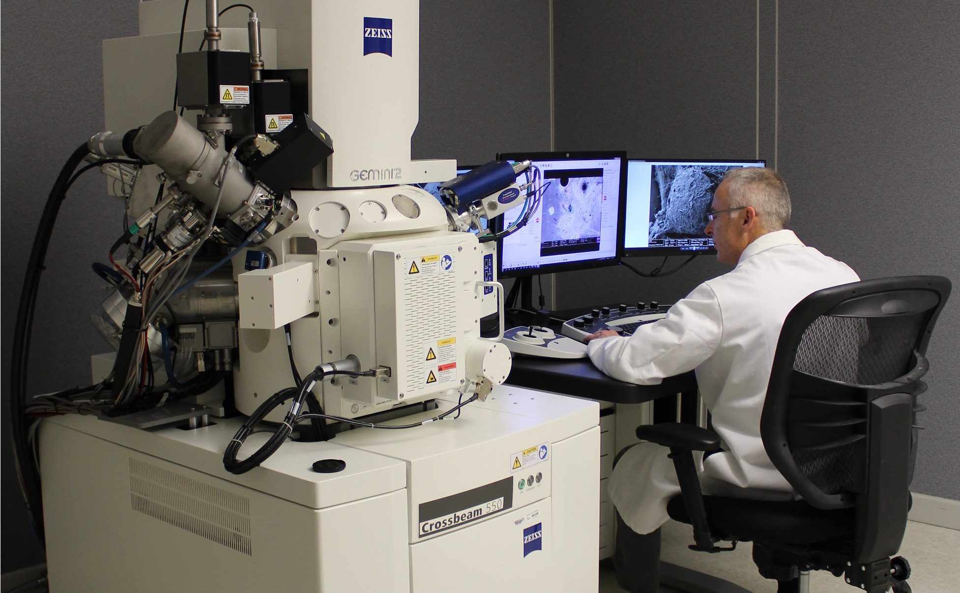
At CvEMC we have acquired a Zeiss Xbeam 550 FIB-SEM and a Zeiss Gemini 450 SEM as well as a Talos L120C TEM. For correlative microscopy we have a Zeiss LSM upright microscope equipped with an AiryScan detector. We also have a full suite of EM preparation equipment including a Leica EM ICE high-pressure freezer and freeze-substitution unit, ASP™-1000 mPrep automatic specimen processor, and a RMC Boeckeler ATUMtome. Segmentation and visualization is done at our workstations equipped with Wacom tablets, and all of our computational work (AI model training etc) is done either at Biowulf or the ATRF compute cluster.
Technology Development
Sample Preparation
We have developed protocols to render challenging samples amenable to vEM imaging. Recently we created hybrid high-pressure freezing (HPF) and quick freeze substitution (QFS) protocols to massively increase metallization of samples, resulting in better SNR in vEMC images (Belanger S et al, 2022) ; we also created cryoCLEM protocols to trap architectural intermediates in the rapidly developing C. elegans embryo (Chang IY et al 2021). We have acquired instrumentation and are developing protocols to allow quick and consistent processing of larger tissue samples.
Efficient vEM
We use imaging SOPs to rapidly and efficiently image targeted features of interest at high resolution, while also occasionally sampling the larger cellular context. The strategy of high-resolution “ROI imaging” and intermediate resolution “keyframe imaging” was originally developed in collaboration with Zeiss Inc and Fibics Inc, is now a central feature of FIB-SEM tomography acquisition (Narayan K et al 2014). We use a combination of AT and FIB-SEM to target specific regions in large specimens (Castranova D et al, 2025).
Correlation
Combining multiple imaging modalities to either relocate or register features of interest is a continuing area of interest in the group. We have standardized protocols that allow for 3D correlative imaging (LM + FIB-SEM) of difficult samples such as dividing cells within the drosophila spermatocytes (Kunduri G et al, 2022) and continue to work on methods that will allow more efficient FIB-SEM imaging of adherent cells. CryoCLEM approaches can be used to trap and image transient events in thick samples by cryo LM and room temperature FIB-SEM (Chang IY et al 2021).
Segmentation & Visualization
This is an area of focus of the lab. We have developed empanada, a plugin on napari for facile training/fine-tuning of deep learning models, as well as running inference and proofreading of model outputs. Still considered a bottleneck in vEMC pipelines, segmentation efforts are now bolstered by our efforts in curating and sharing large datasets of relevant vEM images that greatly increase the performance of pre-trained DL models for downstream tasks such as mitochondrial segmentation (Conrad R and Narayan K 2021). Indeed our generalist model MitoNet is still the state-of-the-art for instance segmentation of mitochondria from 2D and 3D EM image data (Conrad R and Narayan K, 2023). The acceleration of segmentation is now allowing quantitation of features in EM at a scale that allows statistical robustness, such as mitochondria in hepatocytes (Kang SWS et al, 2024), salivary granules (Heydecker M et al, 2024)
Data Handling and Standardization
A community effort to standardize vEMC data and metadata formats is underway. We have contributed to the CLEM and CvEM portions of REMBI metadata recommendations (Sarkans U et al 2021) as well as helped shape community principles for image data sharing (Bajcsy et al, 2025).