Imaging Mass Cytometry Laboratory Workflow
Defined regions of interest are analyzed using the Hyperion XTi™ laser ablation module, which couples high-resolution tissue ablation with time-of-flight mass spectrometry to resolve individual metal isotopes. Isotope signals are computationally integrated to generate highly multiplexed images with single-cell and sub-cellular spatial resolution. Raw data are returned to investigators, with optional downstream analysis including project-specific cell segmentation and quantitative workflows based on the Bodenmiller Lab analysis pipeline.
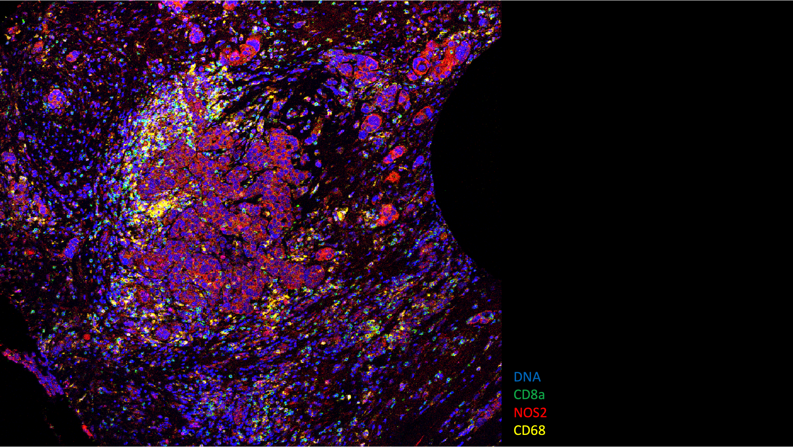
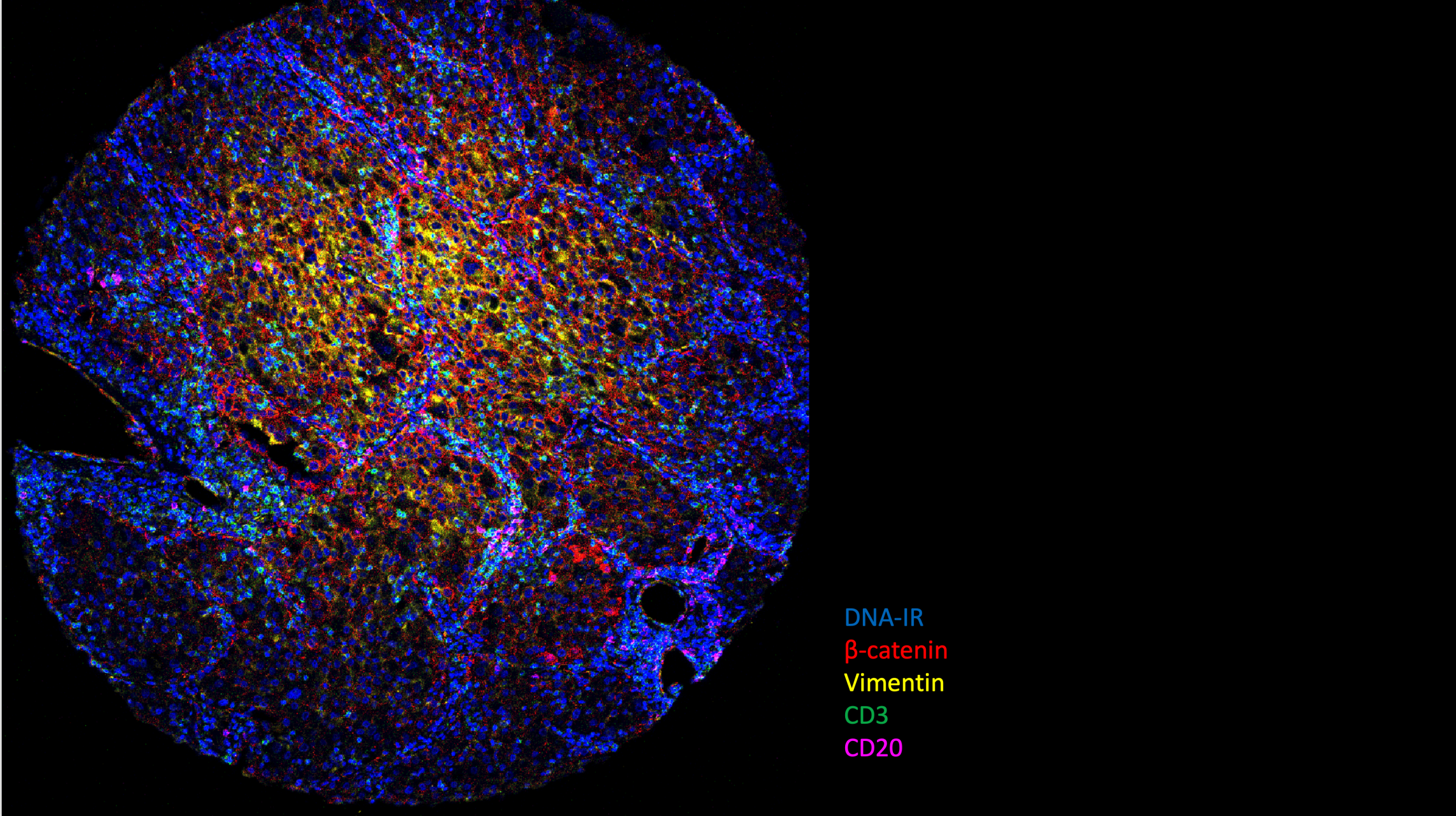
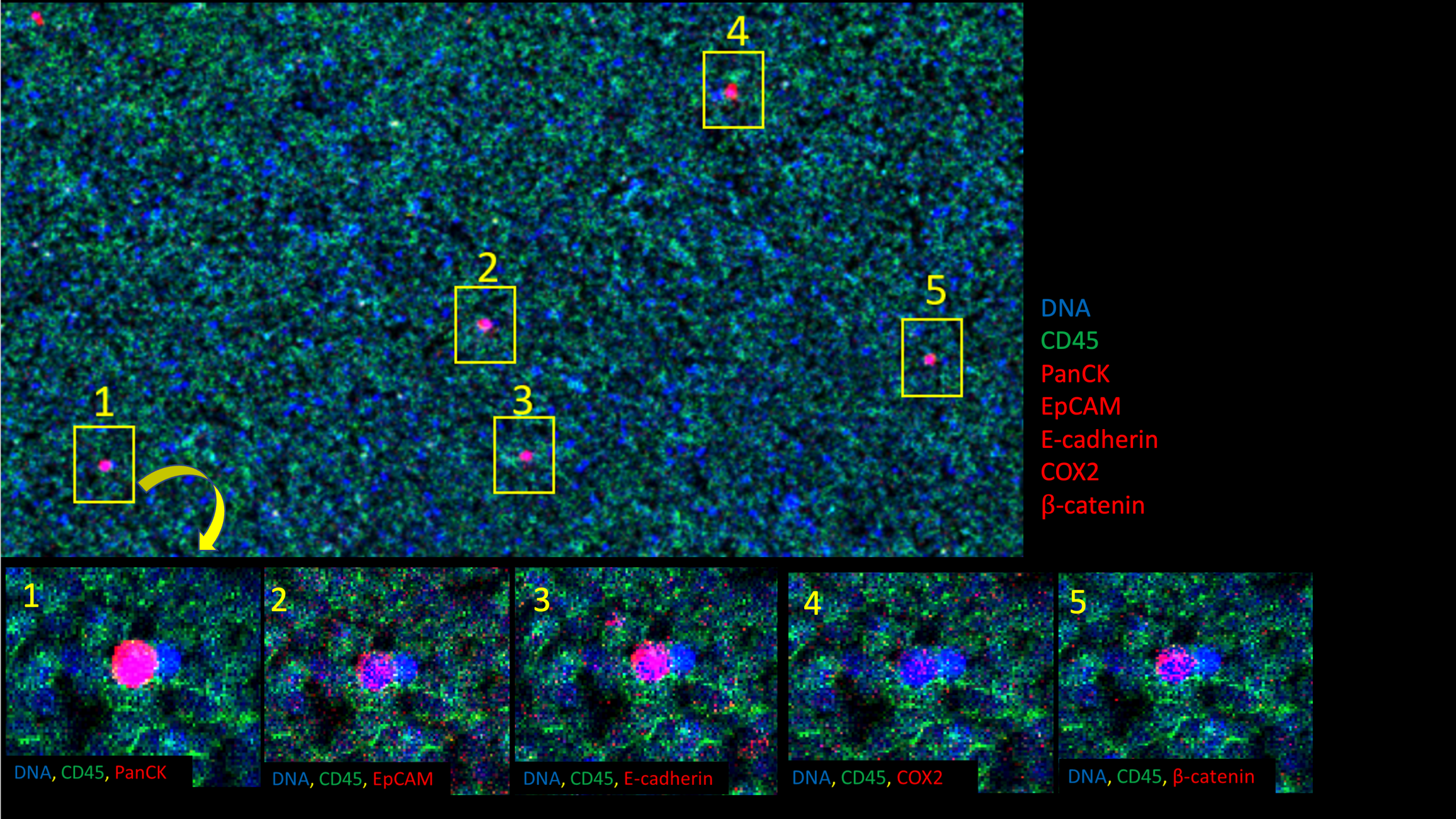
In collaboration with investigators, the MCC designs and validates metal-tagged antibody panels using appropriate positive and negative control tissues or tissue microarrays. Optimized panels are applied to 4–5 µm sections of FFPE, fresh-frozen, or fixed-frozen tissues, as well as cell smears, enabling simultaneous staining of 40+ protein targets while preserving tissue architecture.
Select IMC Projects